
FiTTE has two functions around fertility treatment to solve clinical problems.
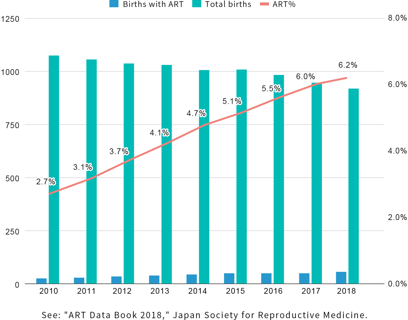
Despite a decrease in the fertility rate in Japan, the number of infants born with assisted reproductive technologies (ART) such as in vitro fertilization(IVF), Intracytoplasmic sperm injection(ICSI), cryopreservation of eggs and embryos, fresh embryo transfer, and frozen embryo transfer is increasing due to technological advances and later marriage. As of 2018, one out of every 16 newborns was ART infants. (Figure below)
In Japan, where fertility treatment technology has advanced, infertility treatment has become so common and it is reported that one in every 5.5 couples experience it. However, infertility treatment is only partially covered by health insurance, and the financial burden on patients has been raised as an important issue.
Preimplantation genetic testing for aneuploidy (PGT-A) is a well-known testing method for embryos chromosomal aneuploidy, which is believed to be a cause of repeated miscarriages.
This method requires invasive procedures on the embryos, and there are concerns about its impact on pregnancy and fertility rates.
In addition, the cost of the procedure tends to be high, placing a heavy burden on the patient.
Based on the method (Ootsuki et al. Fertil Stertil. 2019) which determines the chromosomal aneuploidy non-invasively from the growth process characteristics of embryos (time point identification and area calculation) developed by the clinic and academic partners, NextGeM has developed the image segmentation AI to automatically screen the aneuploidy without human procedures. By conducting a retrospective observational study with approximately one hundred cases and tens of thousands of images in which
time-lapse videos were labeled by physicians and embryologists as training data, an image segmentation AI model was developed and enabled to extract embryo growth process features (time point identification and area calculation).
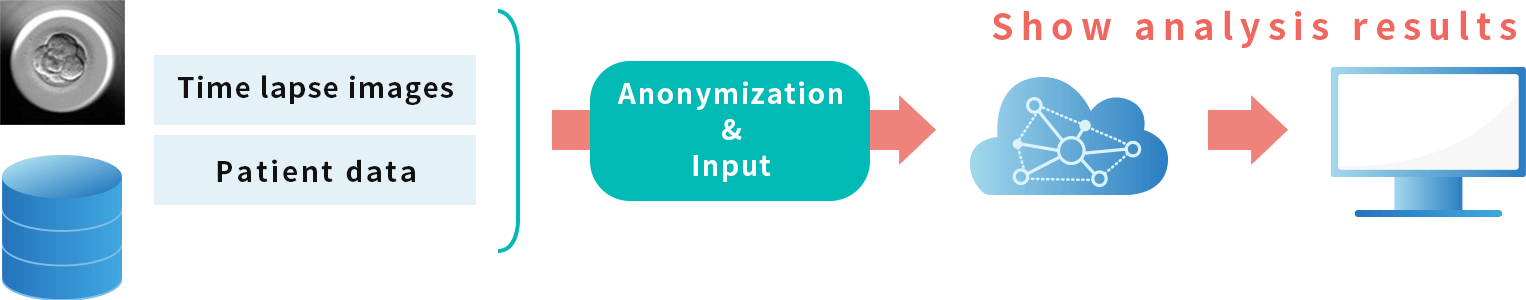
By uploading the time-lapse images of IVF/ICSI embryo culture into the software connected to the image segmentation AI placed on the cloud server, chromosomal aneuploidy determination information is provided to physicians.
This makes it possible to perform inexpensive, non-invasive, and rapid in-clinic testing and provide the necessary information.
(*Physicians should also check other clinical information and make a comprehensive decision.)
*NextGeM is conducting a prospective evaluation study with one of the largest (number of cases) infertility clinics in Japan.
[Supplemental Information]
A mechanism that enables anonymized processing within medical institutions has also been implemented.
A function that allows embryologists to correct the output results by the image segmentation AI is also under development.
Internet connection is required to download and the software can be used for research use.
For more details, please contact us by clicking on the link below
・Citations and references:https://www.fertstert.org/article/S0015-0282(19)30620-X/fulltext
In assisted reproductive technologies (ART), including IVF and ICSI, the condition of the embryo is considerably important for subsequent pregnancy and childbirth, but the current primary method is to visually evaluate the morphology of the embryo at the time of embryo transfer.
Various evaluation methods, such as embryo’s growth process analysis are being discussed at related academic societies, but there is still no established method to predict pregnancy and childbirth.
We have developed an image analysis AI that inputs time-lapse images of the embryo culture process and outputs the results of a five-stage evaluation of pregnancy and childbirth possibility.
As a retrospective observational study of approximately 20,000 cases, the image analysis AI was developed by extracting embryo growth process and morphological characteristics of embryos based on the correlation between time-lapse images of the embryo culture process and pregnancy and childbirth outcomes from the time-lapse images of embryos’ growth process.
(RMB 2022 ”A novel system based on artificial intelligence for predicting blastocyst viability and visualizing the explanation”)
Time-lapse images of IVF/ICSI embryo culture are uploaded into software connected to an image analysis AI placed in the cloud, and the results of a five-stage evaluation can be provided to physicians and
assist in the embryos’ selection for transfer.
・The software will assist physicians in embryo transfer (physicians should also check other clinical information and make a comprehensive decision).
・The image analysis AI model is placed in the cloud and linked to the software.
[Supplemental Information]
This analysis software is for research purposes.
A mechanism that enables anonymized processing data within a medical institution has already been implemented.
The software can be downloaded and used as software within the medical institution. Internet connection is required.
・Citations and references:https://onlinelibrary.wiley.com/doi/10.1002/rmb2.12443